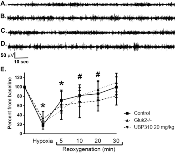
Figure 3.

Effects of hypoxia on background EEG characteristics in control, GluK2−/− and UBP310-treated neonatal mice. (A–D) Representative hippocampal EEG traces at baseline demonstrating background EEG patterns in control mice (A); GluK2−/− mice (B); and control pre- and post-UBP310 injection in the same animal (C and D, respectively). In all groups, background activity consisted of an almost continuous pattern with interspersed sharps and brief periods of low amplitude activity. (E) Exposure to 4% hypoxia for 4 min and 30 sec resulted in significant background attenuation in control (n = 11), UBP310-treated (n = 14) and GluK2−/− mice (n = 8; *p < 0.0001 vs. baseline in all groups, ANOVA). A progressive return to baseline was observed over the first 30 min of reoxygenation. Background activity remained significantly suppressed at 5 min in all groups (*p < 0.0001 vs. baseline in all groups, ANOVA) and at 10 and 20 min after start of reoxygenation in GluK2−/− and UBP310-treated mice (#p < 0.0001 vs. baseline, ANOVA). By 30 min after the start of reoxygenation, background activity was not different than pre-hypoxia. There was no significant difference between groups either with degree of background suppression or timing of return to baseline (p = 0.869, ANOVA). For all analyses of the effects of hypoxia on background EEG activity, sections of EEG without artefacts or seizures were selected at predetermined time points as indicated in the method section. Data shown as percent mean ± SEM.
