Figure 3.
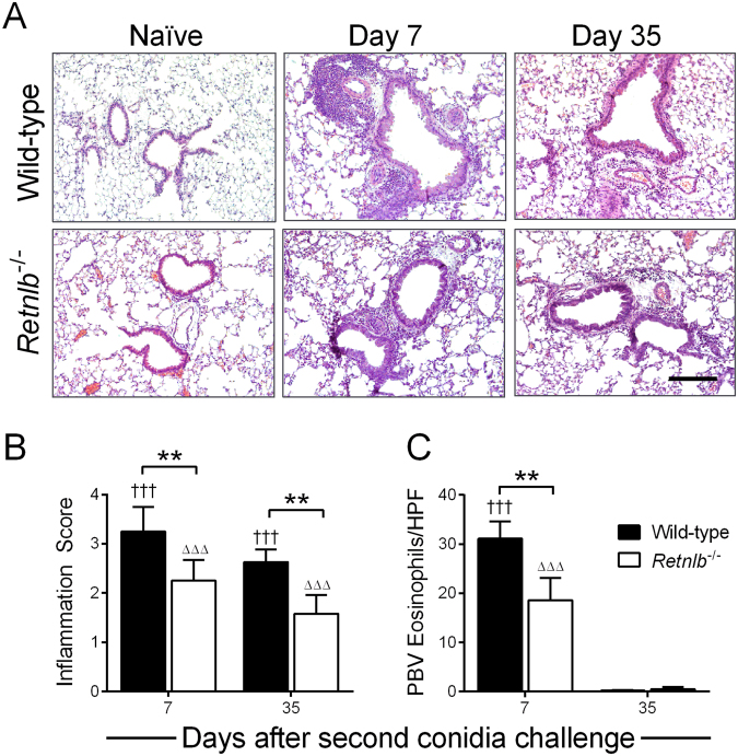
RELM-β promotes peribronchovascular inflammation. Peribronchovascular inflammation was highest at early time points and gradually decreased over time in both genotypes compared to naïve controls (A), and Retnlb−/− mice had less tissue inflammation compared to WT (B). Tissue eosinophils were lower in KO mice compared to WT (C). Scale bar = 200 µm and applicable to all photo micrographs. Naïve mice in each genotype were scored 0 and did not contain tissue eosinophils. Data are represented as the mean and SD of n = 4–7 mice/group in one representative study of two independent studies. Data at each time point were compared by two-way ANOVA with Dunnett’s and Sidak’s multiple comparisons test to their naïve controls († and Δ), and between genotypes (*) respectively. One, two, and three symbols represent P < 0.05, P < 0.01, and P < 0.001 correspondingly. HPF – high power field.

