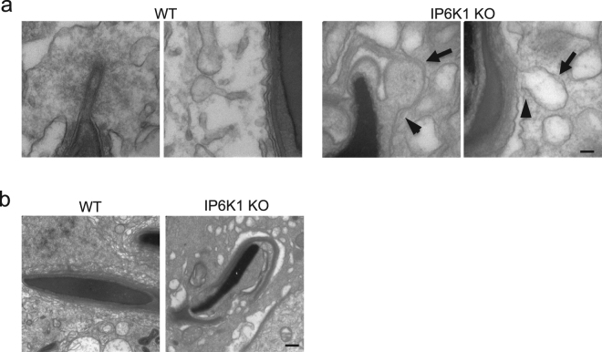
Figure 8.
IP6K1 deletion disrupted spermatid-Sertoli cell interactions. (a) Electron microscopy showed structures of tubulobulbar complexes of WT and IP6K1 KOs. The bulbs were enlarged (arrows) and the tubular regions were absent (arrowheads) in the IP6K1 KOs. Scale bar 100 nm. (b) Electron microscopy showed step 16 spermatids from WT and IP6K1 KO stage VII seminiferous tubules. The KO spermatids showed excess residual cytoplasm and degenerated acrosomes. Scale bar 500 nm.

