Figure 5.
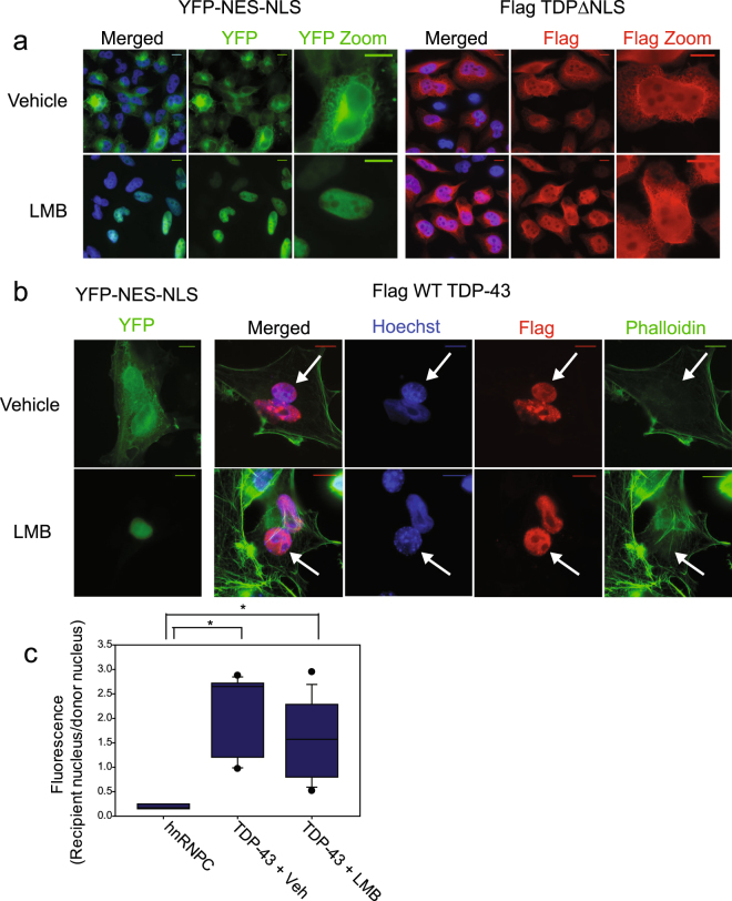
TDP-43 localization and nuclear export is XPO1 independent. (a) Direct fluorescence of HeLa cells expressing fusion protein YFP-NESPKI-NLSSV40 on left. Immunofluorescence using Flag antibody on HeLa Tet-ON cells expressing Flag TDP-43∆NLS on the right. Nuclei were stained using Hoechst. Cells were treated with either Vehicle (Ethanol, −0.1% of total volume) or Leptomycin B (LMB) 10 nM for 12 hours. (b) Left: direct fluorescence of HeLa cells undergoing mock shuttling assay, expressing YFP-NESPKI-NLSSV40. Cells were treated with either Vehicle (Ethanol, −0.1% of total volume) or Leptomycin B (10 nM) for the duration of the assay. Right: Example heterokaryons from shuttling assay. Cells were treated with either Vehicle (Ethanol, −0.1% of total volume) or Leptomycin B (10 nM) for the duration of the assay. Nuclei are stained with Hoechst. Flag WT TDP-43 is detected by immunofluorescence with Flag antibody. Actin cytoskeleton visualized with phalloidin stain. Recipient nucleus (3T3) indicated by arrow. Scale bars- 10 um. (c) Quantification of shuttling assay as in 1b. Two independent experiments. hnRNPC- 3 heterokaryons counted. Flag WT TDP-43, vehicle treated- 8 heterokaryons counted. Leptomycin B treated- 10 heterokaryons counted. * indicates significant difference between groups, p < 0.002 using Mann-Whitney Rank Sum Test. No significant difference between Vehicle and LMB treated heterokaryons, but insufficient power to detect a difference.

