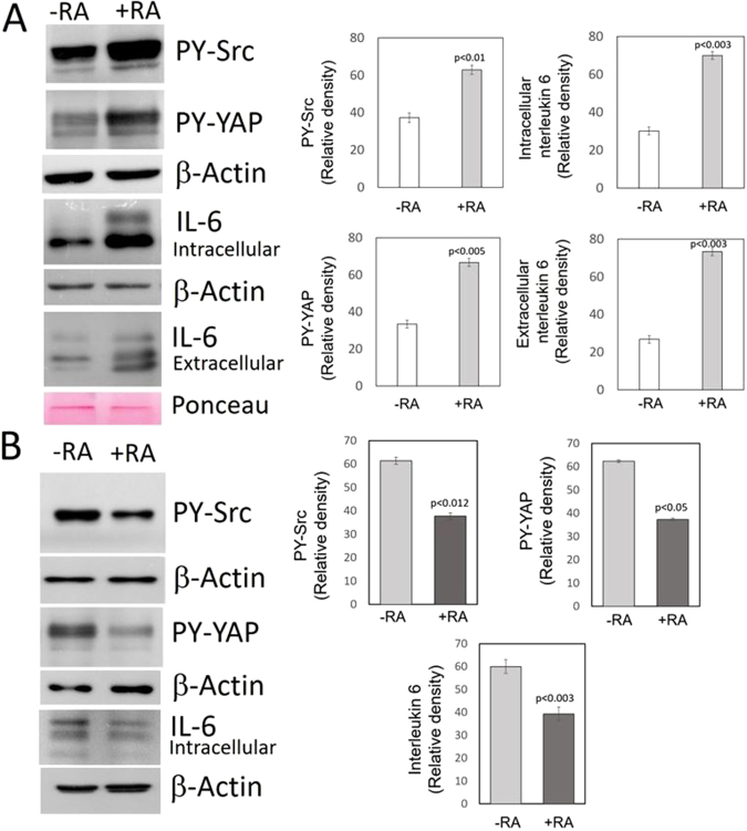
Figure 1.
Effect of RA on the Src-YAP-IL6 axis in triple-negative MDA-MB-231 and MDA-MB-468 breast cancer cells. MDA-MB-231 and MDA-MB-468 breast cancer cells were incubated for two days in the absence (-RA) or presence (+RA) of retinoic acid (5 μM). (A) Western blots of MDA-MB-231 cells show the increase in tyrosine phosphorylation of Src and YAP determined in nuclear extracts and the increase of IL-6 expression assessed in cell lysates and the culture medium. The bar graphs show quantification of data from three independent experiments. β-Actin and Ponceau staining were used as loading controls. (B) Western blots of MDA-MB-468 breast cancer cells show the decrease in tyrosine phosphorylation of Src and YAP determined in nuclear extracts and the decrease of IL-6 expression assessed in cell lysates. The bar graphs show quantification of data from three independent experiments. β-Actin was used as loading control. Full-length figures of the cropped blots are in Supplementary Figures S1–S4.

