Figure 3.
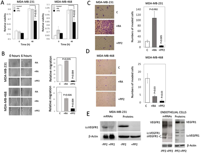
Effect of retinoic acid and the Src inhibitor PP2 on viability, migration, and invasiveness of MDA-MB-231 and MDA-MB-468 breast cancer cells in vitro. Consequences of Src inhibition on the VEGFR1 expression on MDA-MB-231 breast cancer cells and endothelial cells. Prior assessment of viability, migration, and invasion cells were incubated for 48 h in the presence of 5 μM RA or 10 μM PP2. (A) Cell viability of MDA-MB-231 and MDA-MB-468 breast cancer cells incubated in the presence of RA or the Src inhibitor PP2. In both MDA-MB-231 and MDA-MB-468 breast cancer cell lines, cell viability did not change in the presence of RA and decreased upon Src inhibition by PP2. The bar graphs show quantification of data from three independent experiments. (B) Effect of retinoic acid and Src inhibitor PP2 on the migration of MDA-MB-231 and MDA-MB-468 breast cancer cells in vitro. The cell layer of breast cancer cells treated with either RA or PP2 was streaked with a sterile pipet tip to assess migration, and the wound was allowed to recover for 6 hours. Following recovery, the remaining uncovered wound area was measured, and relative migration was calculated as the percentage of wound area covered by cells. In MDA-MB-231 cells, migration increased by retinoic acid and decreased markedly upon Src inhibition. In MDA-MB-468 cells, migration decreased by retinoic acid and decreased markedly upon Src inhibition. The bar graphs show quantification of data from three independent experiments. (C and D) Effect of RA and the Src inhibitor PP2 on invasiveness of MDA-MB-231 and MDA-MB-468 cells in vitro. Breast cancer cells were treated with either RA or PP2, seeded on polycarbonate filters coated with Matrigel, as described in the Materials and Methods Section, and incubated for 24 h. Quantification of invaded cells represents the mean number of cells per field counting seven random fields at 40× or 20× magnification. Treatment with 5 μM RA markedly increased cell invasion in MDA-MB-231 cells (P < 0.0001 vs. control cells) while a decrease was observed in MDA-MB-468 cells. Treatment with the Src inhibitor PP2 (10μM) suppressed cell invasion in both types of cells. E) Consequences of Src inhibition on the VEGFR1 expression in MDA-MB-231 breast cancer cells and endothelial cells. Cells were incubated in the absence (−) or presence (+) of the Src inhibitor PP2. In MDA-MB-231 breast cancer cells, Northern blot analyses show that the expression of the pro-invasive intracellular isoform i21VEGFR1 decreases upon Src inhibition. Western blot analyses show that the protein i21VEGFR1 decreases after Src inhibition. In endothelial cells (HUVEC), Northern blot analyses show that the expression of all VEGFR1 isoforms decreases upon Src inhibition. Western blot analyses show that all the isoforms detected by the antibody against the tyrosine domain of VEGFR1 decrease after Src inhibition. β-Actin was used as loading control. Full-length figures of the cropped blots are in Supplementary Figures S16–S19.

