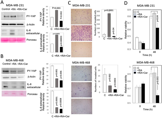
Figure 4.
Effect of RA and RA plus cerivastatin on nuclear YAP, IL-6 expression, and invasiveness in vitro of triple-negative MDA-MB-231 and MDA-MB-468 breast cancer cells. MDA-MB-231 and MDA-MB-468 breast cancer cells were incubated for two days in the presence (+RA) of retinoic acid (5 μM) or the presence of RA (5 μM) plus cerivastatin (1 μM) (RA + Cer). (A) Western blots of nuclear extracts and culture medium of MDA-MB-231 cells treated with RA show the increase of nuclear tyrosine phosphorylated YAP and extracellular IL-6. These actions were reverted when cells were treated with cerivastatin plus retinoic acid. The bar graphs show quantification of data from three independent experiments. β-Actin and Ponceau staining were used as loading control. (B) Western blots of nuclear extracts and culture medium of MDA-MB-468 cells treated with RA show the decrease of nuclear PY-YAP and extracellular IL-6. No major additional changes were observed when cells were treated with cerivastatin plus retinoic acid. The bar graphs show quantification of data from three independent experiments. β-Actin and Ponceau staining were used as loading controls. The density of PY-YAP was normalized to the amount of β-Actin. (C) To assess the effect of RA and RA + cerivastatin on invasiveness of MDA-MB-231 and MDA-MB-468 cells in vitro, treated breast cancer cells were seeded on polycarbonate filters coated with Matrigel, as described in the Materials and Methods Section, and incubated for 24 h. Quantification of invaded cells represents the mean number of cells per field counting seven random fields at 40× or 20× magnification. Treatment with 5 μM RA markedly increased cell invasion in MDA-MB-231 cells (P < 0.0001 vs. control cells) while a decrease was observed in MDA-MB-468 cells. Treatment with RA (5μM) plus cerivastatin (1μM) suppressed cell invasion in both types of cells. (D) Cell viability of MDA-MB-231 and MDA-MB-468 breast cancer cells incubated in the presence of RA (5 μM) or RA (5 μM) plus cerivastatin (1 μM). Cells were incubated for 48 h in the presence of RA or RA plus cerivastatin. In both MDA-MB-231 and MDA-MB-468 breast cancer cell lines, cell viability did not change in the presence of RA and decreased upon incubation in the presence of RA plus cerivastatin. The bar graphs show quantification of data from three independent experiments. Full-length figures of the cropped blots are in Supplementary Figures S20–S21.

