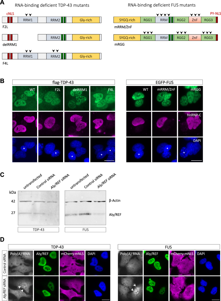
Figure 3.
Nuclear export of TDP-43 and FUS does not require RNA-binding and is independent of the mRNA export machinery. (A) Schematic diagram of RNA-binding deficient TDP-43 and FUS mutants. 2FL = F147L/F149L; 4FL = F147L/F149L/F229L/F231L; mRRM/mZnF = 7 point mutations in the RRM domain/6 point mutations in the zinc finger (see methods for details); mRGG = all Rs in RGG motifs were exchanged for K. (B) The indicated flag-tagged TDP-43 or EGFP-FUS constructs were transiently transfected into HeLa cells and nuclear export was examined in the interspecies heterokaryon assay. Heterokaryons were incubated for 2 h (TDP-43) and 3 h (FUS), respectively, in the presence of cycloheximide and localization of TDP-43 or FUS proteins were visualized by flag immunostaining or direct EGFP fluorescence (green), respectively, hnRNP-C immunostaining (magenta) and DAPI (blue). Both wild-type (WT) and RNA-binding deficient mutant versions of TDP-43 and FUS shuttle from human to mouse nuclei (marked with an asterisk in the DAPI channel). Scale bars: 20 μm. (C) and (D) HeLa cells stably expressing mCherry-TDP-43-mNLS or mCherry-FUS-mNLS were transfected with control or Aly/REF-specific siRNA. 3 days post-transfection, Aly/REF levels in total cell lysates were analyzed by Western blotting, β-actin served as a loading control (C). In parallel, cells were processed for fluorescence in situ hybridization (FISH) and immunocytochemistry to visualize poly(A) + mRNA (white), Aly/REF (green), mCherry-TDP-43/FUS-mNLS (magenta) and were stained with DAPI (blue) (D). Cells with reduced Aly/REF levels show a nuclear accumulation of poly(A) + mRNA (arrows), however no nuclear accumulation of mCherry-tagged NLS mutant TDP-43 or FUS. Scale bars: 20 μm.

