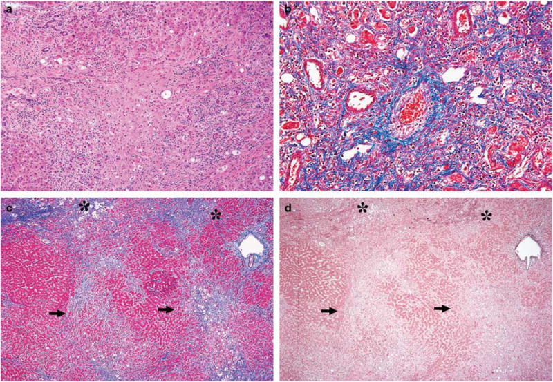
Figure 2.

Additional examples of fibrosis in aggressive non-alcoholic steatohepatitis. (a) Broad dense centrizonal fibrosis, H&E stain. (b) Perivenular sclerosis and intimal thickening. Centrizonal arteries are also present to the left of the vein, trichrome stain. (c) Subacute changes with two-tone trichrome staining; dense, thick collagen bundles stain dark blue (asterisks), while regions with subacute dropout stain pale blue (arrows), trichrome stain. (d) Elastic fiber deposition in early fibrosis (arrows—lack of elastic fiber deposition in regions of necrosis; asterisks—elastic fiber deposition in early fibrosis), orcein stain. (Original magnification: (a): × 100; B: × 200; (c, d): × 40). H&E, hematoxylin and eosin.
