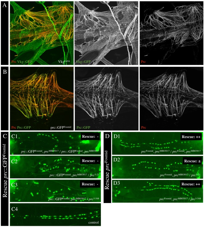
Fig. 3.
Prc::GFP localises to the cardiac ECM but fails to rescue the prc mutations. (A) The cardiac extracellular matrix of a dissected third instar wild-type larva (heart chamber region) stained for the Viking::GFP (anti-GFP, green channel) and endogenously expressed Pericardin protein (anti-Prc, red channel). Viking (Collagen IV) stains all basal laminae whereas Pericardin is restricted to the cardiac matrix. (B) The cardiac extracellular matrix of a dissected third instar wild-type larva (heart chamber region) stained for the Pericardin::GFP fusion protein expressed from the fosmid insertion (anti-GFP, green channel) and endogenously expressed Pericardin protein (anti-Prc, red channel). By staining of the endogenous Prc with anti-Prc, the antibody recognises the Prc::GFP protein as well, which indicates complete overlap. (C) Recombinant prc::GFPFosmid, prcMB03017 animals (C1) were mated with prc3-21 and prc3-548. F1 larvae at the third instar larval stage were analysed for partial or complete rescue of the cardiac phenotype (dissociation of pericardial cells as a read out for cardiac ECM disassembly). The analysis was carried out in homozygous animals with the genotype prc::GFPFosmid, prcMB03017/prc::GFPFosmid, prcMB03017 (C1) and animals with the genotypes prc::GFPFosmid, prcMB03017/prc3-21 (C2) and prc::GFPFosmid, prcMB03017/prc3-548 (C3). (D) prcFosmid was used instead of prc::GFPFosmid for the rescue experiment (D1-D3).

