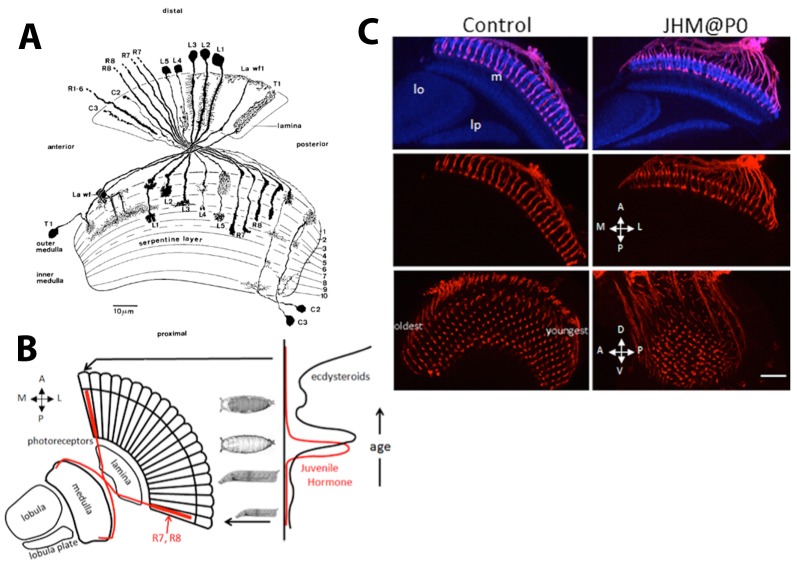
Fig. 1.
Diagram of neurons in the outer optic lobe and the effect of JHM on photoreceptor ingrowth. (A) Camera lucida drawings of the photoreceptor axons (R1-R8) and the neurons innervating the lamina and the medulla of the adult optic lobe of D. melanogaster. See text for the description of the various neurons. (B) Diagram illustrates the progressive ingrowth of R7 and R8 into the medulla of the optic lobe over time at the onset of metamorphosis when the ecdysteroid and JH titers are fluctuating. (C) Effect of JHM on the ingrowth of R7 and R8 photoreceptors into the medulla. The JHM pyriproxifen was applied at the time of pupariation (P0) and the optic lobes of uneclosed day 1 adults were imaged (n=16). The controls were untreated day 1 adults (n=4). The images are sections viewed along the dorsoventral (D,V) axis (top and middle) and along the medial-lateral (M,L) axis (bottom). Red, chaoptin; blue, N-cadherin. m, medulla; lo, lobula; lp, lobula plate. Scale bar: 50 µm. Fig. 1A is Fig. 3A in Fischbach and Dittrich (1989) in Cell and Tissue Research, reprinted by permission from Nature/Springer/Palgrave.

