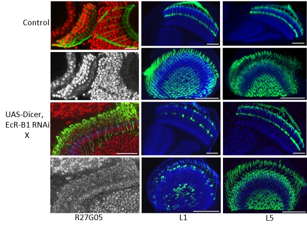
Fig. 7.
The effect of the suppression of EcR-B1 on the development of lamina neurons L1 and L5. The left-hand column shows the myristylated GFP expression of the pan-lamina lines R27G05-GAL4 (Control) and R27G05-GAL4>UAS-Dicer; EcR-B1 RNAi at 39 and 30 h respectively after pupariation together with the gray scale images of the EcR-B1 immunostaining in each. Images typical of three animals each. The next two columns are images of L1 (SS00691) and L5 (SS00692) neurons respectively in normal day 1 adults (top two rows) or in day 1 adults in which Dicer; EcR-B1 RNAi had been expressed at 29°C from the onset of wandering to eclosion (bottom two rows). Images typical of nine animals for L1 and 6 for L5. Images in the left-hand column and in the first and third rows are sections viewed along the dorsoventral axis and those in the second and fourth rows are viewed along the medial-lateral axis. Red, EcR-B1; blue, N-cadherin. Scale bar: 50 µm (25 µm for the R27G05, EcR-B1 RNAi at 30 h APF).

