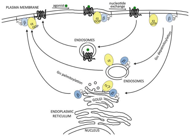
Fig. 11.3. Model of activation-induced G protein trafficking.
Upon GPCR-mediated activation of an appropriate heterotrimeric G protein at the cytoplasmic surface of the PM, different, but not necessarily mutually exclusive, pathways of reversible translocation have been described. One series of studies has shown that after GPCR activation select Gβγ can undergo rapid (within seconds) movement from the PM to the Golgi. Then, Gβγ can return from the Golgi to the PM with similar rapidity. This cycle of Gβγ movement has been described as diffusion-mediated. Interestingly, the rapid activation-induced Gβγ PM-Golgi cycle depends upon palmitoylation, and likely by extension depalmitoylation. However, Gβγ appears to undergo this cycle while Gα remains on the PM; thus, Gα at the Golgi during activation-induced rapid translocation is indicated by the question mark. In another pathway, GPCR-mediated activation promotes depalmitoylation of Gα allowing Gα to translocate off of the PM. This pathway is slower (minutes) than the PM-Golgi pathway. Gβγ has also been observed to show such slower movement off of PM along with Gα. Gα may simply be released into the cytoplasm by virtue of its depalmitoylation, or it may follow a vesicle-mediated pathway involving recycling endosomes together with Gβγ. In this slower pathway, Gα and Gβγ would return to the PM even slower (0.5–1 h). The figure also indicates that Gα and Gβγ traffic independently of GPCR internalization. The models in this figure focus on translocation of G proteins in non-visual systems. See the text and accompanying references for more details regarding light-activated translocation of visual G proteins

