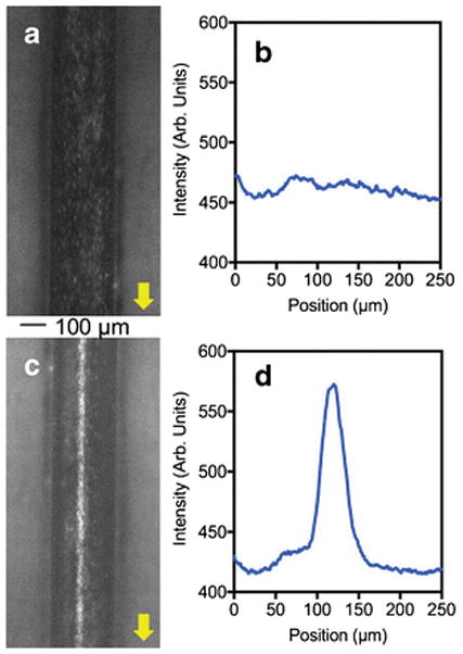
Fig. 5.

Epi-fluorescence micrographs and the plot profile image scanning showing the acoustic focusing of bovine red blood cells in micromachined Al device. Nile red stained bovine RBCs in the absence of acoustic force (a) and its plot profile image scanning obtained from ImageJ software (b). The focused bovine RBCs in the presence of resonance acoustic waves (c) and its plot profile image scanning (d).
