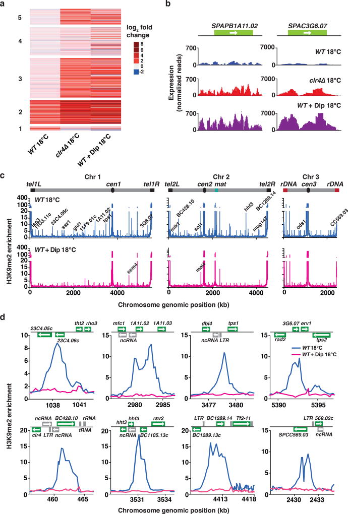
Figure 7. Depletion of iron from growth medium abolishes heterochromatin assembly at low temperature.
(a) Heat map of fold change values for up-regulated transcripts relative to WT 30°C cells in the indicated strains grown at 18°C. Clusters are grouped according to the analysis presented in Fig. 1f. (b) RNA-seq analysis of the expression levels of two representative heterochromatic loci are shown for the indicated strains grown at 18°C. (c) ChIP-chip analysis of genome-wide H3K9me relative enrichment in WT cells at 18°C, either untreated or treated with 250µM Dip. New 18°C facultative heterochromatin peaks are indicated on the WT 18°C plot. Note that H3K9me at constitutive heterochromatin domains (cen, mat and tel) and meiotic islands (ssm4 and mei4) is largely maintained in Dip-treated cells. WT (untreated) ChIP-chip data is also presented in Fig. 2a. (d) H3K9me relative enrichment at individual loci in WT cells, either untreated or treated with 250µM Dip, at 18°C. WT (untreated) ChIP-chip data is also presented in Fig. 2b. Source data for Fig. 7a are available with the paper online.

