
Fig. 5. Organoids remodel their matrix microenvironment in a PKD-dependent manner.
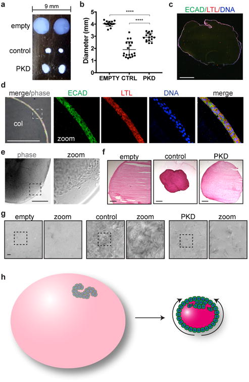
a, Photograph of organoids implanted into collagen balls and cultured in suspension for two weeks. b, Diameters of empty (n=17) and organoid-implanted (ctrl, n=19; PKD, n=16) collagen droplets, pooled from three experiments (empty vs. ctrl, t(21.96)=13.53, p=3.9334×10⁻¹²; ctrl vs. PKD, t(24.74)=11.33, p=1.7989×10⁻⁶). Each droplet is indicated by a single data point. c, Wide-field immunofluorescence image of whole droplet compacted by a PKD organoid, stained for tubule segment markers. Zoom of dashed boxed region is shown for fluorescent channels. d, Confocal immunofluorescence at the edge of a representative droplet after contraction, adjacent to the collagen interior (col). The dashed boxed region is shown for each individual channel at higher magnification. e, Phase contrast images showing the edge of a representative droplet at an early stage of compaction. f, Sirius red staining of collagen droplets. g, TEM 25,000X images of collagen filament structure in the interior of droplets. Zoom of dashed boxed region is shown for each image. h, Schematic of collagen droplet compaction by organoids. KTECs migrate out and surround the scaffold, which contracts towards the outgrowth (curved arrows). Scale bars, 500 μm (b-f) or 500 nm (g).