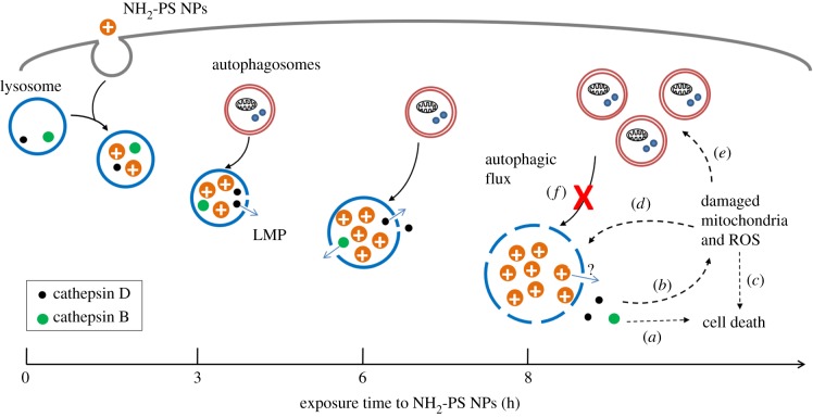
Figure 5.
Scheme of LMP and deregulated autophagy induced by NH2-PS NPs. NH2-PS NPs are endocytosed into lysosomes. After 3 h, these NPs cause mild LMP, leading to leakage of small lysosomal components (processed cathepsin D, 27 kDa) into cytosol. Gradually, after 6 h, the LMP is exacerbated, marked by the release of larger lysosomal components (cathepsin B, 38 kDa). Lysosomal expansion due to the ‘proton sponge' effect of these cationic NPs can be also observed. At this point, the NP-loaded lysosomes might still be functional, and autophagosomes generated from basal level autophagy can still be fused and degraded. However, after 8 h exposure to NPs, lysosomal expansion and LMP continue to aggravate. The released cathepsins can directly lead to caspase-independent cell death (process a), or perturb mitochondria (process b), resulting into ROS generation and apoptotic cell death (process c). The generated ROS can lead to more severe LMP, serving as a feedback loop (process d). Autophagy is induced, likely due to the damaged mitochondria and ROS (process e). However, the generated autophagosomes cannot be fused with and/or degraded by lysosomes, as they are extensively damaged due to accumulation of NH2-PS NPs (process f).

