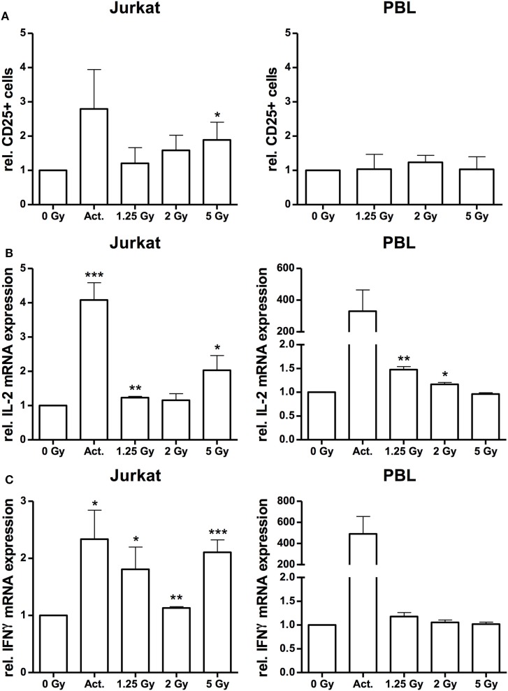
Figure 6.
Irradiation stimulates immune activation in Jurkat cells and peripheral blood lymphocytes (PBL). FACS analysis of CD25 surface expression on Jurkat cells and CD3-positive PBL (A) following irradiation with a dose of 1.25, 2, and 5 Gy. Stimulation with 25 µl/ml CD3/CD28/CD2 T-cell activator (Act.) in Jurkat cells or mock-irradiated cells served as controls (N = 3). In PBLs, an activator could not be applied due to inference with the CD3 stimulus. Quantification of interleukin (IL)-2 (B) and interferon-γ (IFNγ) (C) mRNA expression by quantitative real-time PCR in Jurkat cells and PBL at 24 h after irradiation with a dose of 1.25, 2, or 5 Gy. Stimulation with 25 µl/ml CD3/CD28/CD2 T-cell activator (Act.) or mock-irradiated cells served as controls (N = 2). Data are represented as mean + SD. Student’s t-test compared activator-treated or irradiated cells with non-irradiated controls; *P < 0.05, **P < 0.01, ***P < 0.001.

