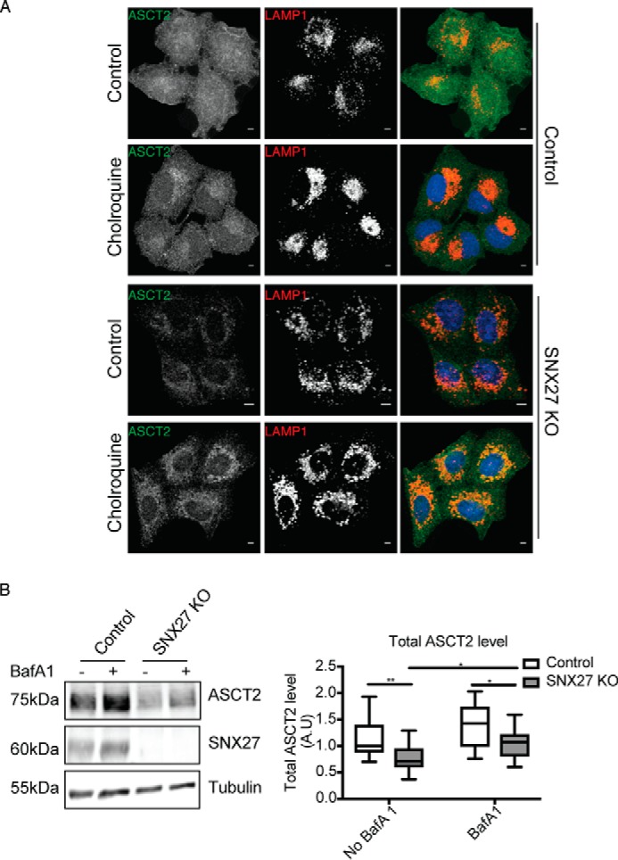
Figure 3.

Knockout of SNX27 mis-sorts ASCT2 for lysosomal degradation. A, HeLa control and SNX27 KO cells were left untreated or treated with 50 μm chloroquine for 16 h before fixation and immunolabeling with ASCT2 and LAMP1 antibodies. Scale bars = 5 μm. B, HeLa control and SNX27 KO cells were left untreated or treated with 200 nm Bafilomycin A1 for 10 h in the presence of 100 μg/ml cycloheximide. Equal numbers of HeLa control and SNX27 KO HeLa cells were subjected to Western blotting and stained with antibodies against ASCT2, SNX27, and tubulin. Representative blots from three independent experiments are shown, and the calculated molecular weight for each protein is indicated. The -fold differences for ASCT2 between HeLa cells and SNX27 KO HeLa cells are presented (means ± S.D.). Two-tailed Student's t test indicates the difference between HeLa parental and SNX27 KO cells. **, p < 0.01, HeLa versus SNX27 KO cells, no Bafilomycin A1; *, p < 0.05, HeLa versus SNX27 KO cells, Bafilomycin A1–treated; *, p < 0.05, untreated SNX27 KO versus Bafilomycin A1–treated SNX27 KO cells.
