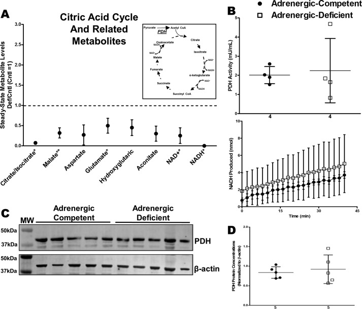
Figure 9.
Effects of adrenergic deficiency on PDH activity and protein concentrations. A, steady-state metabolites involved in TCA cycle in E11.5 adrenergic hormone-deficient hearts compared with controls (dotted line). Schematic representation of TCA cycle and related pathways provided as reference. B, PDH activity in adrenergic hormone-competent (●) and adrenergic hormone-deficient (□) embryonic hearts, expressed as milliunits of enzyme per ml of sample and NADH produced per min, respectively. Milliunits/ml are calculated as ((nmol of NADH)/(min)(ml of sample)). C and D, PDH protein concentrations (39 kDa) in adrenergic hormone-deficient embryonic trunks (□) compared with controls (●), normalized to β-actin. The β-actin panel is re-used as a representative image of six replicated experiments probing for GAPDH, G-6-PDH, and PDH. Numerical values below the x axes refer to the number (n) of samples analyzed. Student's t test was used to compare means between competent and deficient groups. *, p < 0.05; **, p < 0.01.

