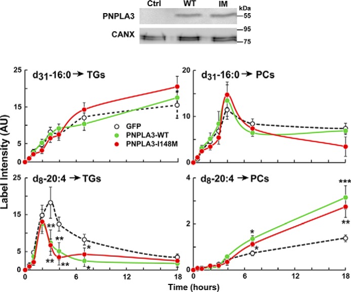
Figure 8.

Synthesis of TG and PL in in HuH7 cells overexpressing PNPLA3. HuH7 cells were infected with a GFP adenoviruses (GFP, dotted line), PNPLA3(WT) (green), or PNPLA3(148M) (red). After 2 days, palmitate-d31 and arachidonate-d8 were conjugated with BSA and added to the medium at a final concentration of 1 μm each. Cells were then harvested at the indicated time points (n = 3 dishes/time point). Expression of PNPLA3 constructs was analyzed by immunoblotting using a V5 antibody. Lipids were extracted, and the incorporation of the isotope-labeled FAs into TGs and PCs was measured by direct infusion lipidomics. PCs were identified by the 184-Da MS2 fragment in positive mode. TGs were identified based on their FA neutral loss. Values were normalized to the PC and TG standards in the SPLASH standard mixture. The experiment was repeated twice with similar results. Error bars, S.D. *, p < 0.05; **, p < 0.01; ***, p < 0.001.
