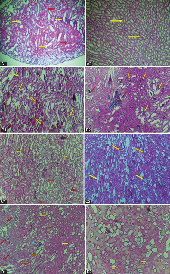
Figure 3.

Light microscopic evaluation of the renal tissues stained with hematoxylin and eosin. (a1 ×20) and (a2) Normal histology of the kidney tissues, showing normal glomerulus (red arrow) and normal tubules (yellow arrow) (Group A). (b1) and (b2) Kidney sections of Group B, showing glomerular deformation (red arrow), tubular dilatation, vacuolation, swelling, and degeneration of their lined epithelial cells (yellow arrows), vascular congestion (orange arrow), inflammatory cell infiltration (blue arrow), and fibrinoid dystrophy (green triangle) (×20). (c1, c2) and (d1, d2) Kidney sections of Group C and Group D, respectively, showing the enhancement in tubular and glomerular injuries and other pathologic alterations. The red star represents the hyaline dystrophy (×20).
