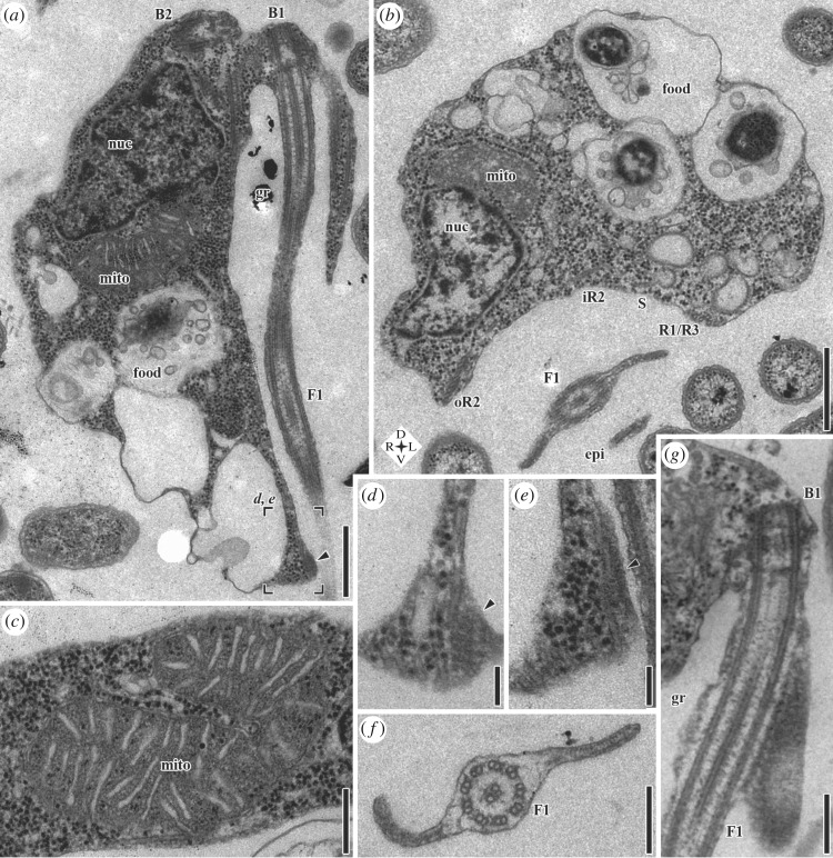
Figure 2.
Transmission electron microscopy (TEM) of G. okellyi. (a) Longitudinal section; note marks for position of images d and e, detailing composite fibre (arrowhead). (b) Transverse section, showing architecture of ventral feeding groove; note compass rose for orientation. (c) Mitochondrion, showing discoidal cristae in transverse section. (d,e) Serial sections of posterior part of right margin of groove in same cell as a, showing striated and dense portions of composite fibre (arrowhead), respectively. (f) Transverse section of posterior flagellum (F1), showing vanes. (g) Longitudinal section of proximal portion of posterior flagellum, showing origin of dorsal vane, and striations of its lamella. B1, basal body 1 (of posterior flagellum); B2, basal body 2 (of anterior flagellum); epi, epipodium; F1, posterior flagellum; food, food vacuole; gr, groove; iR2, inner portion of microtubular root 2; mito, mitochondrion; nuc, nucleus; oR2, outer portion of microtubular root 2; R1, microtubular root 1; R3, microtubular root 3; S, singlet microtubular root. Scale bars: (a) 500 nm, (b) 500 nm, (c) 250 nm, (d) 100 nm, (e) 100 nm, (f) 200 nm, (g) 250 nm.

