Figure 3.
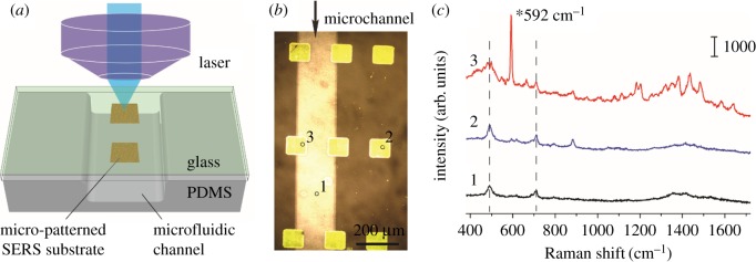
Raman spectra of Nile blue measured at different positions of the microfluidic SERS chip: (a) illustration of SERS detection at the SERS-active substrate inside a microfluidic channel; (b) an optical microscopic image of the SERS-active substrate (yellow squares) embedded in a microfluidic chip; and (c) three SERS spectra measured at three representative positions of the microchannel (b), 592 cm−1 peak is the characteristic peak of Nile blue. SERS were excited by a 785 nm laser with a laser power of approximately 3 mW and acquisition time of 10 s.

