Figure 20.

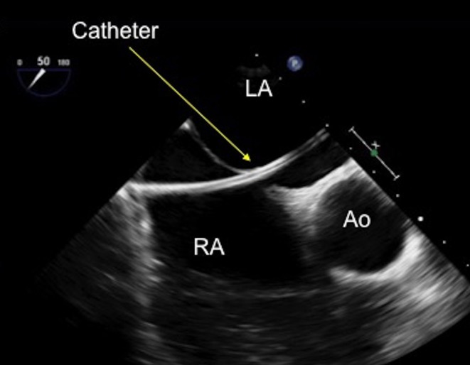
TOE image at 50° mid-oesophageal view with a catheter seen clearly crossing the defect and advanced into the mid-left atrium. Ao, aorta; LA, left atrium; RA, right atrium.

TOE image at 50° mid-oesophageal view with a catheter seen clearly crossing the defect and advanced into the mid-left atrium. Ao, aorta; LA, left atrium; RA, right atrium.