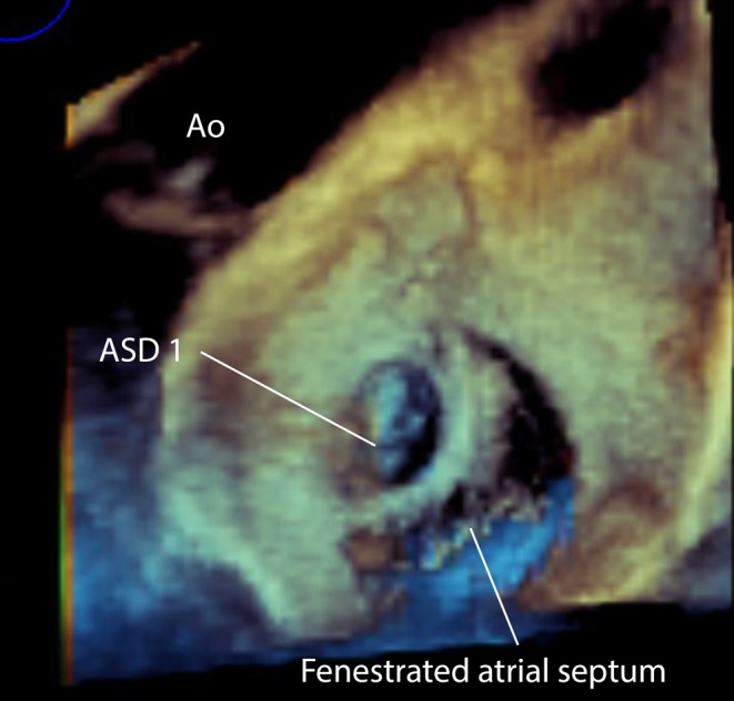
Figure 16.

3D TOE image at mid-oesophageal level viewed from the left atrial aspect showing a small superior secundum atrial septal defect and a larger mutlifenestrated atrial septal defect. This has implications for the planning of device closure as two devices may be required to close the defects. ASD, atrial septal defect.

 This work is licensed under a