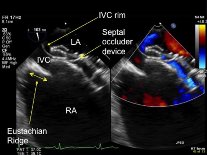
Figure 18.

TOE image at 100° low oesophageal view showing a prominent Eustachian ridge (see double-ended arrow), which has the potential to be confused with the IVC rim. In this image the IVC rim has been well caught by the septal occluder device. IVC, inferior vena cava; LA, left atrium; RA, right atrium.

 This work is licensed under a