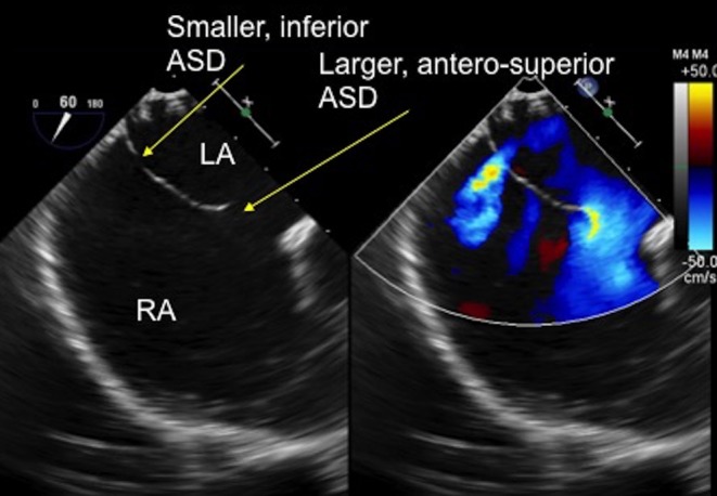
Figure 19.

TOE image at 60° mid-oesophageal level showing two separate defects in the thin atrial septal tissue. There is a larger, antero-superior defect with a small aortic rim and a smaller, more inferior defect which are clearly seen with the colour Doppler. ASD, atrial septal defect; LA, left atrium; RA, right atrium.

 This work is licensed under a