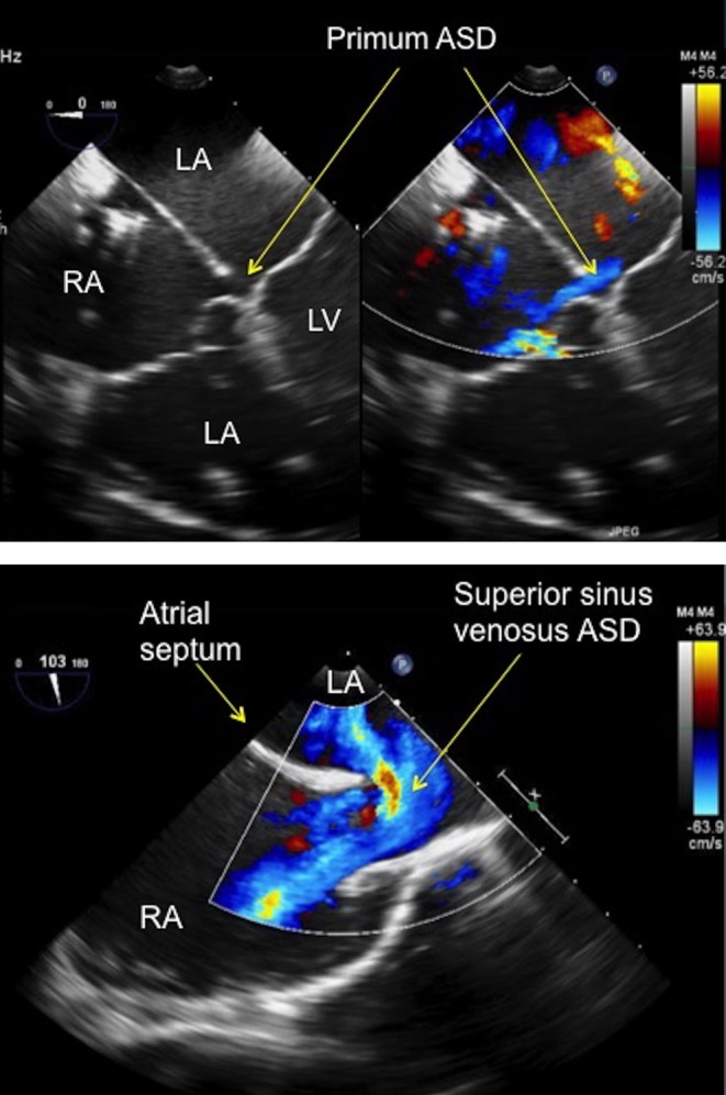
Figure 2.

(A) TOE image at 0° mid-oesophageal level four-chamber view showing a small primum atrial septal defect (see arrows) in a patient with a partial atrioventricular septal defect. The defect has no rim to the atrioventricular valve, which precludes percutaneous device closure. ASD, atrial septal defect; LA, left atrium; LV, left ventricle; RA, right atrium; RV, right ventricle. (B) TOE image at 100°, high oesophageal level showing a large superior sinus venosus atrial septal defect with left to right shunt seen on colour Doppler. Note that there is no rim to the superior vena cava. ASD, atrial septum; LA, left atrium; RA, right atrium.

 This work is licensed under a