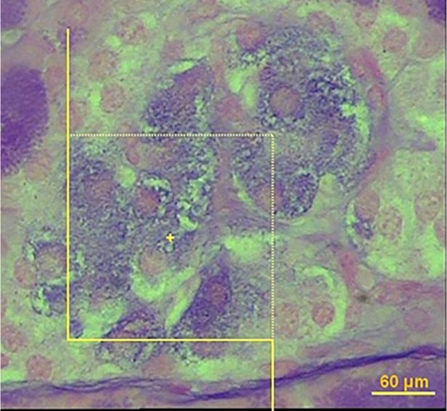
Figure 3.
Estimation of the number of β-cell using the optical disector. The nucleoli profiles of the β-cell were counted, in solvent-treated diabetic group (A), Amygdalus lycioides (1000 mg/kg) extract-treated group (B), and insulin-treated diabetic group (C). The nucleoli were counted only if they were inside or partially inside the sampling frame and none of their parts touched the exclusion lines of the frame (40X, aldehyde fushin staining

