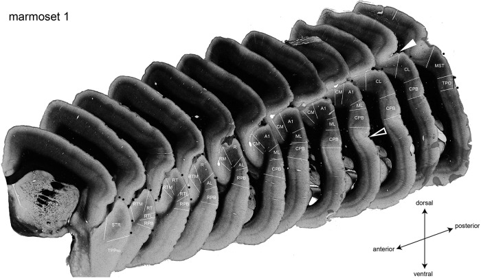
Figure 1.
Coronal sections from the left hemisphere of a marmoset stained for myelin and the areal demarcation of the auditory cortical areas. Auditory cortical areas are defined by the myelin structure. The white lines indicate the areal borders. The black filled circles represent positions of blood vessels. The filled and open white arrowheads indicate the lateral sulcus and the superior temporal sulcus, respectively.

