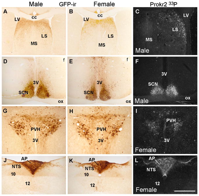
Fig. 2.
Distribution of Prokr2-Cre GFP neurons and Prokr2 mRNA in male and female brain (G3 generation, P60–80 days of age). A–L Bright- and dark-field images showing distribution of GFP immunoreactive cells (GFP-ir) and Prokr2 mRNA in male (A, C, D, F, G, J) and female (B, E, G, I–K) brains. 3V third ventricle, 10 motor nucleus of the vagus nerve, 12 hypoglossal nucleus, AP area postrema, cc corpus callosum, f fornix, LS lateral septum, LV lateral ventricle, MS medial septum, NTS nucleus of the solitary tract, ox optic chiasm, PVH paraventricular nucleus of the hypothalamus, SCN suprachiasmatic nucleus. Scale bar A, B 400 μm, C–L 200 μm

