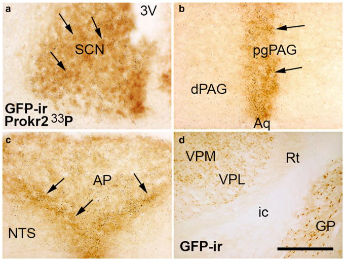
Fig. 4.
Coexpression of Prokr2 mRNA and Prokr2-Cre GFP in the female brain. a–c Bright field images showing colocalization of Prokr2 mRNA (hybridization signal visualized as dark grains) and Prokr2-Cre GFP immunoreactivity (GFP-ir, arrows) in the suprachiasmatic nucleus (SCN, a), in the pleioglial periaqueductal gray (pgPAG, b) and in the nucleus of the solitary tract (NTS, c). d Bright field images showing GFP immunoreactive cells in the ventral posterolateral (VPL) and ventral posteromedial (VPM) nuclei of the thalamus. 3V third ventricle, AP area postrema, Aq aqueduct, dPAG dorsal column of the periaqueductal gray, GP globus pallidus, ic internal capsule, ox optic chiasm, Rt reticular nucleus of the thalamus. Scale bar a–d 100 μm

