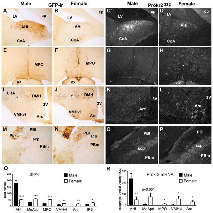
Fig. 5.
Differential expression of Prokr2-Cre GFP and Prokr2 mRNA in adult (P60-P80, G2 generation) male and female brains. A, B, D, E, G, H, J, K Bright- and dark-field images showing the differential distribution of GFP immunoreactive (GFP-ir) cells and Prokr2 mRNA in male (A, C, E, G, I, K, M, O) and female (B, D, F, H, J, L, N, P) brains. Q Bar graphs showing sexual dimorphism in the number of GFP-ir cells in amigdalo-hippocampal nucleus (AHi, P = 0.0007, q = 0.00036, t 6.34), posterodorsal subdivision of the medial nucleus of the amygdala (MeApd, P = 0.003, q = 0.00079, t 4.40), medial preoptic area (MPO, P = 0.0006, q = 0.00068, t 8.38), in the arcuate nucleus (Arc, P = 0.0015, q = 0.0005, t 5.03), in the ventrolateral subdivision of the ventromedial nucleus of the hypothalamus (VMHvl, P = 0.0048, q = 0.0009, t 4.03) and in the lateral parabrachial nucleus (PBl, P = 0.02, q = 0.0043, t 2.82). R Bar graphs showing sexual dimorphism of Prokr2 mRNA in AHi (P = 0.0079, q = 0.040, t 4.91), in MPO (P = 0.036, q = 0.045, t 2.68), in VMHvl (P = 0.020, q = 0.045, t 3.13), in Arc (P = 0.029, q = 0.045, t 2.83) and a strong trend in MeApd (P = 0.051, q = 0.052, t 2.41). No difference was observed in Prokr2 mRNA in the PBl. 3V third ventricle, CoA cortical nucleus of the amygdala, cp cerebral peduncle, DMH dorsomedial nucleus of the hypothalamus, f fornix, LHA lateral hypothalamic area, LV lateral ventricle, ox optic chiasm, PBm medial parabrachial nucleus, Pur Purkinje cells, scp superior cerebellar peduncle. *P<0.05; **P<0.01; ***P<0.001, ****P<0.0001. Scale bar 400 μm

