Fig. 6.
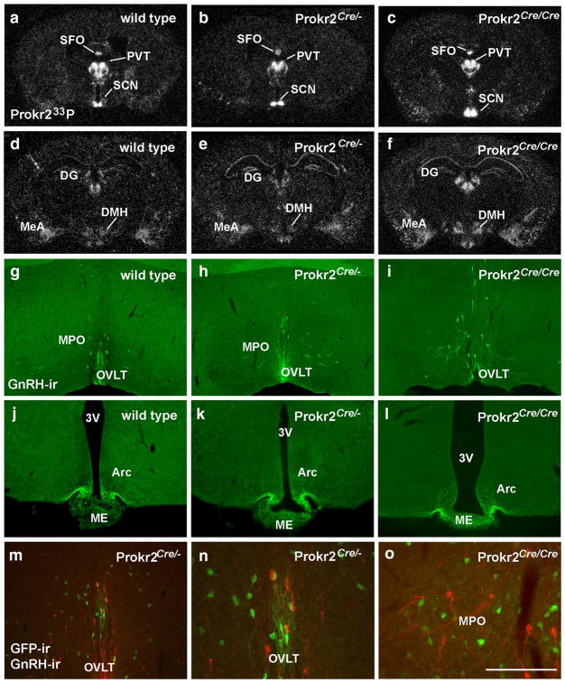
Prokr2 mRNA and GnRH neuronal migration are not altered in Prokr2-Cre heterozygous and homozygous mice. a–f Dark field images showing the distribution of Prokr2 mRNA (hybridization signal observed as silver grains) in the brains of wild type (a, d), Prokr2-Cre heterozygous (b, e G3 generation, P60–80 days of age) and Prokr2-Cre homozygous (c, f G5 generation, P60–70 days of age) females. g–l Fluorescent images showing the distribution of gonadotropin releasing hormone immunoreactive (GnRH-ir) cell bodies (g–i) and fibers (j–l) in wild type (g, j), Prokr2-Cre heterozygous (h, k), and ProR2-Cre homozygous (i, l) female mice. No differences were observed in the distribution of Prokr2 mRNA and GnRH neurons. m–o Fluorescent images showing distribution of GnRH-ir (red) and GFP-ir (green) cells in the Prokr2-Cre heterozygous (m, n) and homozygous (o) female mice. Note lack of colocalization and close proximity between these two groups of cells. 3V third ventricle, ac anterior commissure, Arc arcuate nucleus, DG dentate gyrus, DMH dorsomedial nucleus of the hypothalamus, ME median eminence, MeA medial nucleus of the amygdala, MPO medial preoptic area, OVLT vascular organ of the lamina terminalis, PVT paraventricular nucleus of the thalamus, SCN suprachiasmatic nucleus, SFO subfornical area. Scale bar a–f 2 mm, g–m 400 μm, n, o 200 μm

