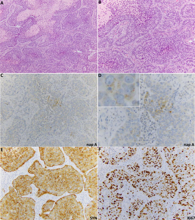
Figure 1.

Example of LCNEC with focal napsin A expression (case ID 16 in Table 2). (A) H&E sections illustrate classic LCNEC morphology, including nested growth pattern with peripheral nuclear palisading, frequent rosette-like arrangements, and areas of geographic necrosis. (B) Higher-power image illustrates non-small cell cytomorphology - moderate volume of cytoplasm and evident nucleoli. Panels C and D illustrate weak and focal but convincing granular cytoplasmic napsin A labeling. Panel E illustrates diffuse labeling for synaptophysin. F. Ki67 marker confirms high proliferation rate (70%).
