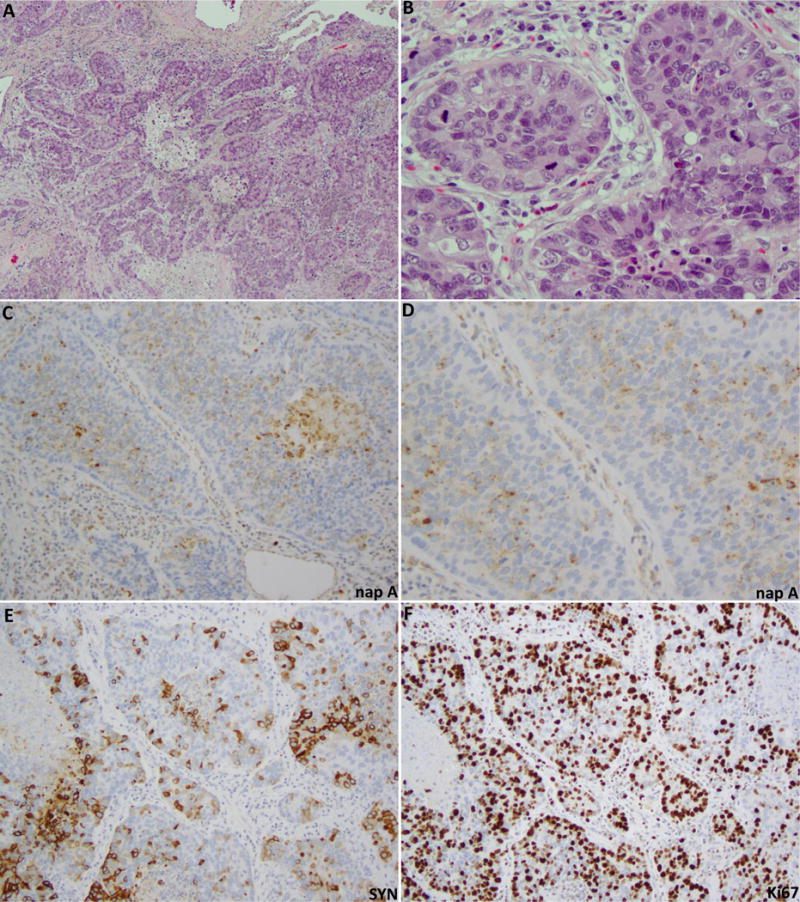
Figure 2.

Example of LCNEC with diffuse weak to moderate napsin A expression (case ID 4 in Table 2). H&E sections (A,B) illustrate neuroendocrine morphology - nested and trabecular growth pattern with peripheral nuclear palisading, and overtly non-small cell cytology - prominent nucleoli with moderate amount of cytoplasm. Tumor has amphophilic cytoplasmic commonly seen in LCNEC. This type of morphology enters in the differential diagnosis with solid adenocarcinoma. Panels C and D illustrate napsin A labeling in the majority of tumor cells, which shows typical granular cytoplasmic reactivity with variably-sized granules. Napsin A expression is seen in the absence of entrapped pneumocytes or histiocytes, confirming the specificity of labelling. Panel E illustrates focal labeling for synaptophysin (SYN) in the same tumor areas as those labeling for napsin A. F. Ki67 confirms high proliferation rate (80%).
