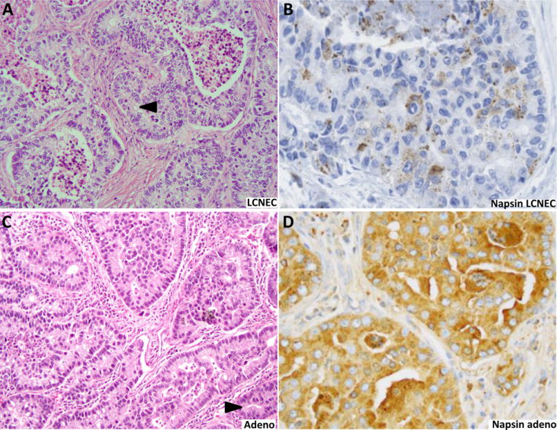
Figure 3.

Contrast in intensity of napsin A reactivity in LCNEC (A and B) and lung adenocarcinoma (C and D). Intense (3+) labeling typical of adenocarcinomas (D) was not seen in any LCNECs (B). The figure illustrates a case on adenocarcinoma with cribriform pattern, which enters in the close differential diagnosis with LCNEC. Cribriform spaces in adenocarcinoma tend to have more undulating outlines, with occasional slit-like lumens, whereas luminal borders in LCNEC are characteristically rosette-like, with rigid/punched-out outlines (arrowheads). Spaces in rosettes also tend to be smaller, pinpoint-like, compared to more variable luminal sizes in adenocarcinoma.
