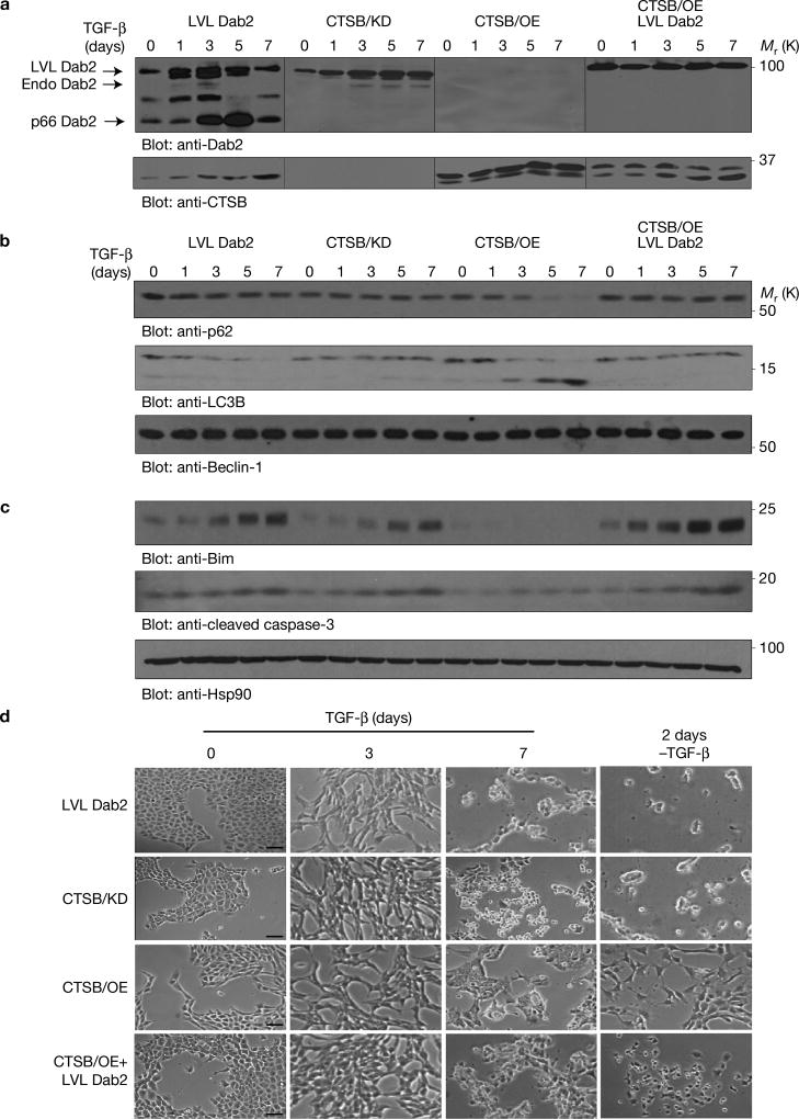
Figure 3.
CTSB-mediated Dab2 degradation promotes TGF-β-induced autophagy. (a) NMuMG cells and their derivatives were treated with TGF-β for the times indicated before the preparation of whole-cell lysates and immunoblot analyses using anti-Dab2 and anti-CTSB antibodies. The resultant images are the product of time average data. The lines on the western blots demarcate individual blots that were run in parallel. (b) The same as in a, but with anti-p62, anti-LC3B and anti-Beclin-1 antibodies. (c) The same as in a, but with anti-Bim, anti-cleaved caspase-3 and anti-Hsp90 antibodies. Hsp90 expression served as a loading control. (d) Morphological analysis of modified cell lines after 3 days and 7 days of TGF-β treatment and 2 days after the removal of TGF-β from cells treated for 7 days with TGF-β. Scale bars, 100 µm. The experiments were repeated three times and similar results were observed. Unprocessed original scans of blots are available in Supplementary Fig. 9.

