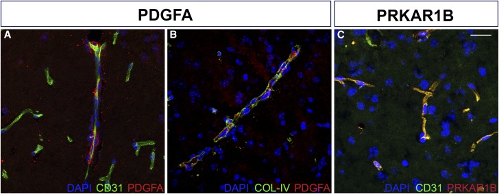
Figure 6.
PDGFA and PRKAR1B localize to vascular structures in the mouse brain. PDGFA (red) shows close localization to (A) endothelial cells (CD31) and (B) basement membrane (COL-IV), components of the vascular substructure. (C) PRKAR1B (red) shows punctate expression in the region of blood vessels (CD31, green). See Materials and Methods for antibody details. Bar, 20 μm.

