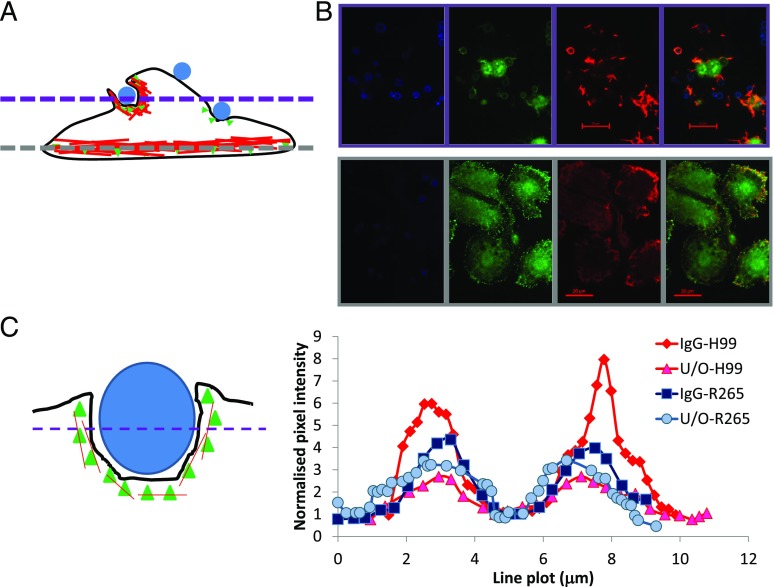
FIGURE 3.
Activated Syk is essential for the uptake of Cryptococcus particles. Mouse macrophage cell line J774.A1 was challenged with either (IgG-opsonized or unopsonized, U/O) C. neoformans H99 or C. gattii R265 for 15 min (B), processed for immunofluorescence, and analyzed by confocal microscopy of localized phospho-Syk (B and C) as described in Materials and Methods. (A) Schematic diagram J774.A1 macrophage with intracellular actin cytoskeleton (red) and yeast particles (blue). To confirm phospho-Syk localization, the bottom of the cells was observed first [(A), grey dashed line and (B), bottom panels], before moving to the middle of the cells [(A), purple dashed line, (B), top panels]. Pixel intensities for 20 cells per sample were determined [(C), right] and normalized to the intensity at the center of the cell [(C), left]. (A and C) The green triangles denote phospho-Syk. The black line denotes the outline of a cell as imagined from the side (i.e., its z-axis). Results are expressed as the mean ± SD of at least three independent experiments. Scale bar, 20 μm.

