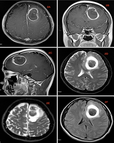
Figure 2.

MRI image done at our facility. A-C) are axial, coronal and sagittal T1 image respectively. D,E) are axial T2 images while 2F is FLAIR. These images show ring like lesions that are consistent with Glioblastoma. The delay window of the evolution process was up to 14 days.
