Figure 6.
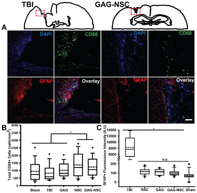
Activated macrophage and reactive astrocyte presence surrounding the lesion site in TBI only control and CS-GAG-NSC treated animals. (A) Representative images of the region corresponding to the red dotted box surrounding the lesion area in coronal brain sections obtained from TBI only control, and CS-GAG-NSC treated animals. Cellular nuclei are represented by DAPI (blue); activated macrophages are represented by CD68 labeled cells (green); and reactive astrocytes are represented by GFAP labeled cells (red). Merged overlays are presented in the bottom right panels in each group. (B) A significantly greater CD68+ reactivity was observed in brain sections obtained from animals treated with NSCs only, and with CS-GAG-NSCs when compared to all other groups. (C) Brain tissue obtained from TBI only controls indicated a significantly increased GFAP immunoreactivity for reactive astrocytes when compared to all treatment groups and sham control. Statistical significance is represented by ‘*’ which indicates p < 0.05. The lack of statistical significance between groups is denoted by ‘n.s’. Scale =100 μm.

