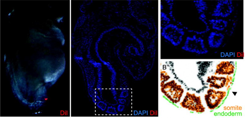
Fig. 1. Example of a DiI labeled Embryo.

A) A 7-somite (S) embryo labeled over the second somite on the right side with DiI (red). B) A DAPI stained sagittal section of the same embryo shows DiI (red) in the pre-pancreatic endoderm and in the underlying somitic mesoderm, better seen in enlargement (B′), and site of DiI release (arrowhead). C) False color rendition of the same section highlights endoderm (green) and somites (orange). Hf, head fold; h, heart; the somites are numbered.
