Figure 1.

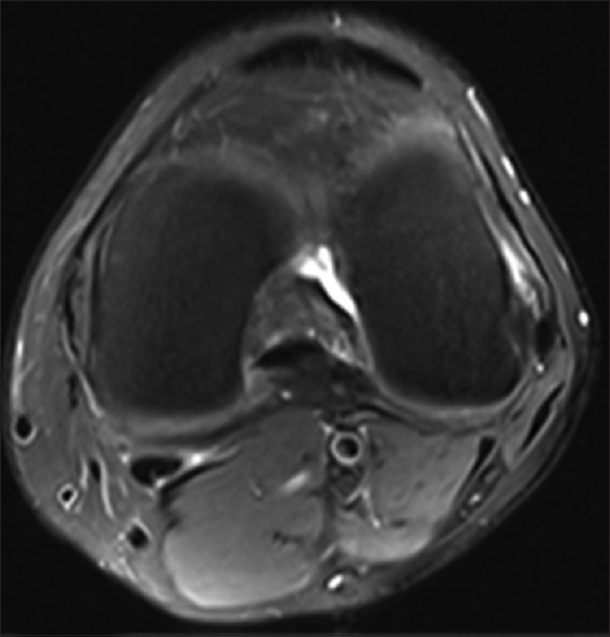
A 47-year-old healthy control. Axial PD-weighted MRI image with fat suppression of the left knee. MRI: Magnetic resonance imaging; PD: Proton density.

A 47-year-old healthy control. Axial PD-weighted MRI image with fat suppression of the left knee. MRI: Magnetic resonance imaging; PD: Proton density.