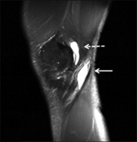
Figure 3.

A 50-year-old AAIHP. Sagittal PD-weighted MRI image with fat suppression, showing a fluid-filled SM-TCL bursa (white dashed arrow) and popliteal bursa (white solid arrow). PD: Proton density; AAIHP: Asymptomatic amateur ice hockey players; MRI: Magnetic resonance imaging; SM-TCL: Semimembranosus-tibial collateral ligament.
