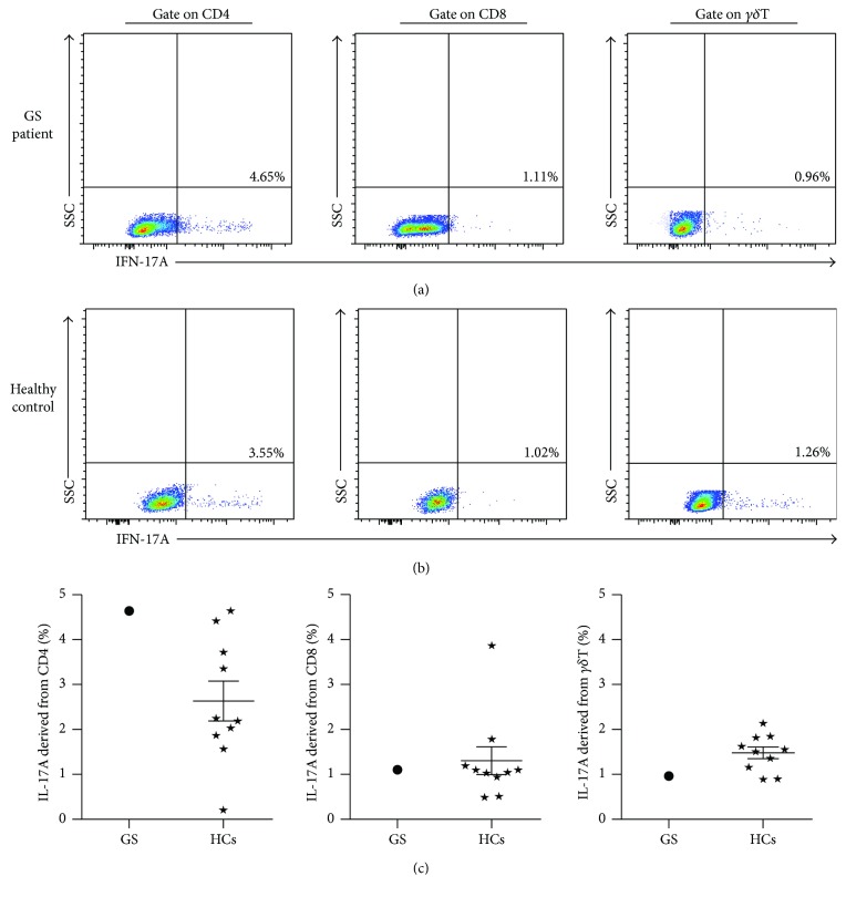
Figure 8.
Cellular IL-17A levels derived from circuiting immune cells in this GS patient and HCs. Representative dot pots of IL-17A derived from CD4+ T cell, CD8+ T cell, and γδT cell in this GS patient (a) and HCs (b). Statistical graphs of intracellular IL-17A in GS patient and HCs (c; N = 10, resp.). ⋆: HCs group.

