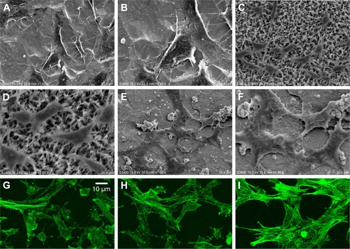
Figure 10.
SEM morphologies of the MC3T3-E1 cells on bare Ti substrate (A and B), HA coating (C and D), and FAgHA coating (E and F); fluorescence microscopic images of MC3T3-E1 cells on the Ti (G), HA (H), and FAgHA (I) coatings after 24 hours of culture, actin were stained with phalloidin (green).
Abbreviations: SEM, scanning electron microscope; HA, hydroxyapatite; FAgHA, F-and-Ag-substituted HA.

