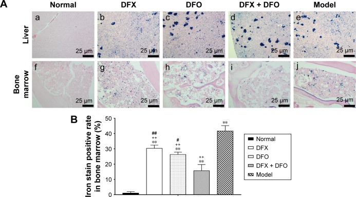
Figure 5.
Iron deposition in liver and bone marrow among different groups.
Notes: (A) Following Prussian blue staining, slides were redyed with Sudan red, observed under a light microscope, and images captured at 400× magnification. Images show (a–e) liver and (f–j) bone marrow, respectively. (B) Data are shown as mean ± SEM (n=3), **P<0.01 (as compared with Normal); ++P<0.01 (as compared with Model); #P<0.05, ##P<0.01 (as compared with DFX + DFO). Normal: Normal control group, DFX: DFX-treated group, DFO: DFO-treated group, DFX + DFO: DFO and DFX cotreated group, Model: Composite model group.
Abbreviations: DFO, deferoxamine; DFX, deferasirox; SEM, standard error of the mean.

