Figure 1.
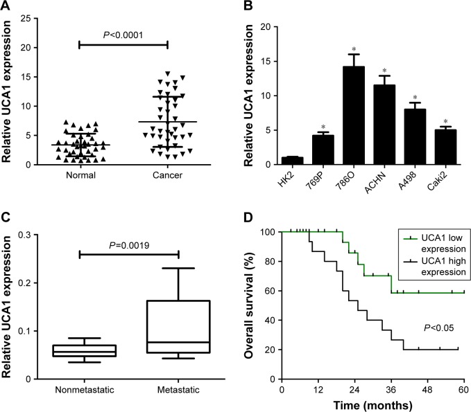
UCA1 was highly expressed in RCC tumor tissue and cells.
Notes: UCA1 expressions in 40 pairs of RCC tumor tissue and adjacent normal tissue (A), RCC cells (769P, 786O, ACHN, A498, Caki2) and normal proximal tubular epithelial cell line (HK2) (B), different pathogenic status (metastatic [n=22] and nonmetastatic [n=18] phases) (C). (D) Kaplan–Meier analysis of overall survival for RCC patients according to differences in UCA1 expression. Median UCA1 expression in RCC tumor tissue was used as a cutoff point to divide the UCA1 high-expression group and the UCA1 low-expression group. *P<0.05.
Abbreviation: RCC, renal cell carcinoma.

